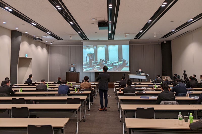
Program
11:30 – 11:35 Opening Remarks
Session 1
Chair: Mitsugu Todo, Research Institute for Applied Mechanics, Kyushu University
11:35 - Presentation 1
Evaluation of bone healing process and bone strength at removal of nail after intramedullary nailing for femoral shaft fracture
Hideyuki Mimata1, 4, Yusuke Matsuura2, Sei Yano2, Seiji Ohtori2, Mitsugu Todo3
1Interdisciplinary Graduate School of Engineering Sciences, Kyushu University, Fukuoka, Japan, 2Department of Orthopeadic Surgery, Graduate School of Medicine, Chiba University, Chiba, Japan, 3Research Institute for Applied Mechanics, Kyushu University, Fukuoka, Japan, 4Research Center of Computational Mechanics, Inc. Tokyo, Japan
For intramedullary (IM) nailing for femoral shaft fracture, no quantitative evaluation method has been established for the progress of bone healing and the decision to remove the nail. We aim to quantify the failure risk of callus and bone strength at removal of nail after IM nailing using quantitative CT-based finite element analysis (QCT/FEA) including callus.
QCT/FEA was performed using CT after IM nailing with joint reaction force and muscle forces at the maximum load during gait. The volume ratio of the interfracture callus with a tensile failure(TF) risk of 100% or more was defined as the TF volume ratio and evaluated. Furthermore, virtual models with the nail removed were created and the bone strength was obtained from the displacement-load curve.
TF volume ratio was 11.6% at 6 months postoperatively, with the failure elements concentrated on the lateral side, and then decreased to 2.6 and 0.5% at 12 and 15 months. It was consistent with radiographically observed osteogenesis, suggesting the possibility of quantitative evaluation of the degree of bone healing. If the cutoff value leading to nonunion can be derived by increasing the number of cases, it is expected to be applied to early judgment of nonunion and pseudarthrosis surgery.
The strength at removal increased and exceeded that of the healthy side at 15 months. It indicated the possibility of clarifying the decision of when to remove the nail by using criteria such as healthy side ratio and life strength.
11:50 - Presentation 2
Mechanical evaluation of screw position of bone fixation device for proximal femur fracture
Yoshihiro Tomita1, Jiro Sakamoto2
1Department of Mechanical Science, Graduate School of Natural Science and Technology, Kanazawa University, Kanazawa, Japan, 2Advanced Manufacturing Technology Institute, Kanazawa, Japan
In this study, we carried out biomechanical analysis to evaluate the stresses on the tip and the screw holes of the device, and examined the screw hole position of the device to consider breakage risk of the gamma nail, an osteosynthesis device for proximal femur, after fracture surgery. A proximal femur fracture model with a complete fracture at the lower part of the trochanter and an incomplete fracture at the trochanter was created, based on the femur model for tutorial MECHANICAL FINDER. Young’s modulus of the fracture region was given a lower value than femur one. Thirteen types of CAD data with different screw hole positions provided by a medical device manufacturer were used for the model of gamma nail. The loading condition assumed a static standing position of one leg with a body weight of 50 kg. Distal surface of the knee joint was completely fixed, and the loading direction was set at the angle recommended for the analysis of the femur in Mechanical Finder. As a contact condition, Coulomb friction was defined at the interface between the femur and the gamma nail, and the friction coefficient of μ = 0.3 was set. Linear elastic analysis was performed on the femur fracture model with the above boundary and contact conditions. At the result, the maximum Mises stress at the screw hole tended to increase as the screw hole position became farther from the top of the nail. Therefore, it is suggested that the closer the screw hole position to the top of the nail makes the broken risk lower.
12:05 - Presentation 3
Investigation about biomechanics of hip fractures using fresh frozen cadavers and finite element analysis
Sei Yano, Yusuke Matsuura
Department of Orthopaedic Surgery, Graduated School of Medicine, Chiba University, Chiba, Japan
We researched about biomechanics of hip fractures using fresh frozen cadavers and finite element analysis.
A total of 26 proximal femurs were retrieved from 16 fresh-frozen cadavers. After CT was taken, mechanical test simulating a sideways fall configuration was performed. The experiment was continued until a hip fracture occurred, and its fracture type (femoral neck fracture or intertrochanteric fracture) was recorded. We simulated two fall patterns (lateral and posterolateral) and examined the effect of fall direction on fracture type. Furthermore, three-dimensional finite element models were created from the CT data, and the reproducibility of the mechanical tests were verified.
In the lateral group, neck fractures and intertrochanteric fractures occurred in equal proportions. Whereas in the posterolateral group, intertrochanteric fractures were occurred in all cases. The concordance rate of fracture type between mechanical testing and FEA was 84.6%.
Luncheon Presentation
12:30 - Presentation 4
Assessing Spinal Strength in Transplant Patients Using 2D and 3D Modeling Approaches
Kent D. Carlson1, Ejigayehu G. Abate2, Hillary W. Garner3, Daniel E. Wessell3, Dan Dragomir-Daescu1
1Department of Physiology and Biomedical Engineering, Mayo Clinic, Rochester, MN, USA, 2Division of Endocrinology, Mayo Clinic Florida, Jacksonville, FL, USA, 3Department of Radiology, Mayo Clinic Florida, Jacksonville, FL, USA
Bone mineral density (BMD) is used for assessment of bone health in the general population. However, BMD alone does not predict the risk of fracture in liver transplant patients. Prevalence of vertebral fracture is estimated to range from 13-56% in pre-transplant subjects with end stage liver disease. We developed a 2D FEA toolkit that uses BMD data from transplant patients to determine vertebral fracture strength of un-fractured vertebrae using compressive bone fracture simulations. We are using this 2D toolkit to estimate vertebral strength in transplant patients, to determine the tool’s ability to predict vertebral fractures. Since no experimental fracture strength data was available for this cohort, we compared the predictions from this 2D toolkit with 3D FEA compressive fracture models developed and simulated with MECHANICAL FINDER. We did this for three patients in the cohort for which we had spinal CT data in addition to BMD scans. We created separate Mechanical Finder models using two different bone material property datasets, to compare these strength estimates with 2D results and with each other. Results from corresponding 2D and 3D simulations were compared. We found significant differences between the 2D and 3D results. These differences require further investigation, and mechanical testing of cadaveric vertebra is needed for model validation.
12:45 - Presentation 5
Computed Tomography-Based Finite Element Analysis for Assessing Skeletal Trauma
Mikoláš Jurda1、Luboš Řehounek2
1Department of Anthropology, Faculty of Science, Masaryk university, Bruno, Czech Republic, 2Department of Mechanics, Faculty of Civil Engineering, Czech Technical University in Prague, Prague, Czech Republic
Finite-element analysis (FEA) is a powerful tool for simulating structural stress analysis of objects based on their shape and defined physical characteristics. The presented study introduces an analysis of CT-derived FEA models for assessing the mechanism of sustained skeletal fractures.
The finite element analysis, represented here by algorithms incorporated in a MECHANICAL FINDER software, was employed to investigate a comminuted fracture of tibia as observed in naturally mummified remains of a 38-year-old man. The association between the trauma-inducing stress and the observed fracture pattern was investigated using an FEA model of the unimpaired right tibia. The mechanical properties were estimated in high resolution by the software based on a voxel density of CT scans. The performed dynamic simulations included low and high-velocity impacts of projectiles and metal splinters of various shapes and dynamic energy.
The performed analysis captured some of the general features of the observed fracture pattern. The outputs were in line with the assumption that the trauma resulted from a high-velocity blunt force impact. While the tools allowed for valuable case-specific simulations, more profound research should determine their robustness to changes in technical factors (e.g., voxel size, physical setup) and their applicability to diverse skeletal parts.
13:00 - Presentation 6
Are internal structures preserved in vertebrates’ fossils useful for Finite element analysis?
– A pilot analysis using multiple preservation fossils–
Kumiko Matsui1, 2
1Department of Paleobiology, National Museum of Natural History, Smithsonian Institution, DC, USA, 2The Kyushu University Museum, Kyushu University, Fukuoka, Japan
Paleontology is one of the study fields that reveal taxonomy, phylogeny, ecology, the evolution of life, and past environments by using fossils. Within these 15 years, FEM in paleontology is very active. FEM allows paleontologists to estimate bite forces of extinct animals quantitively, and these results allow to compare bite forces across taxa. These results provide paleontologists to understand biomechanical understandings and the roles of extinct animals in their ecosystem. On the other hand, fossil-based FEM still has problems. Unlike extant skeletal materials, all fossil materials were damaged and deformed by pressures from surrounded sediments and geo-tectonics between their death and discoveries. In addition to that, almost all internal cavities of fossil skeletons are filled with sediments and minerals. For the above reasons, it is difficult to understand the internal structure and morphology of fossil skeletons correctly, so previous analyses have been limited to simplified models. In this study, to solve these problems, I analyzed multiple X-ray CT datasets of extinct carnivoran specimens from different localities and ages and have various preservation qualities by using both homogeneous and heterogeneous models and compared these results. I will show the results of pilot analyses and want to discuss that.
13:15 – 13:35 Coffee Break
Session 2
Chair: Yusuke Matsuura, Department of Orthopaedic Surgery, Graduated School of Medicine, Chiba University
13:35 - Presentation 7
Cadaveric analysis of acetabular cartilages stress; Comparison of finite element analysis and pressure loading test
Keizo Wada, Yasuaki Tamaki, Tomohiro Goto, Daisuke Hamada, Koichi Sairyo
Department of Orthopaedics, Tokushima University Hospital, Tokushima, Japan
The purpose of this study was to examine the consistency between the finite element analysis and the pressure loading test as a method for evaluating the acetabular joint surface stress.
Four fresh-frozen cadavers with healthy hip were included. The left and right acetabulum and femur were extracted from the cadavers as hip joints. For the acetabulum, the ilium was fixed to the pedestal with plaster. A load of 100 N was applied from the distal part of the femur along the bone axis, and the articular surface pressure was measured with a pressure sensor sandwiched between the acetabulum and the femoral head. Subsequently, the acetabulum was tilted 5 degrees and 10 degrees posteriorly, and a load was applied in the same manner to measure the articular surface pressure. On the other hand, a finite element model was created from the CT image, a cartilage model was added to the head and acetabulum, and finite element analysis was performed under the same conditions to measure the equivalent stress applied to the acetabular joint surface.
When the relationship between the average value of the joint surface pressure by the pressure sensor and the average value of the equivalent stress by the finite element method was evaluated using Pearson’s correlation coefficient, a significant positive correlation was found at r = 0.41.
The results of this study suggest that stress analysis using the finite element method may predict the acetabular joint surface pressure.
13:50 - Presentation 8
Total hip arthroplasty using a three-dimensional porous titanium acetabular cup: an examination of micromotion using subject-specific finite element analysis
Takaki Miyagawa, Haruhiko Akiyama
Department of Orthopaedic Surgery, Gifu University School of Medicine, Gifu, Japan
Background: We investigated the mid-term clinical and radiological results of total hip arthroplasty (THA) using a three-dimensional (3D) porous titanium cup and analyzed the micromotion at the interface of the cup using subject-specific finite element (FE) analysis.
Methods: We evaluated 73 hips of 65 patients (6 men and 59 women; mean age at the time of surgery, 62.2 years; range, 45-86 years) who had undergone THA using a 3D porous titanium cup. We assessed the fixation of the acetabular component based on the presence of radiolucent lines and cup migration using anteroposterior radiographs. Subject-specific FE models were constructed from computed tomography data.
Results: Radiolucent lines were observed in 26 cases (35.6%) and frequently appeared at DeLee and Charnley Zone 3. Following the FE analysis, the micromotion at DeLee and Charnley Zone 3 was significantly larger than that at Zone 2. Furthermore, micromotion was large in the groups in which radiolucent lines appeared at Zone 3.
Conclusions: The mid-term clinical outcome of THA using a 3D porous titanium cup was excellent. However, radiolucent lines frequently appeared at DeLee and Charnley Zone 3. FE analysis indicated that micromotion was large at the same site, strongly suggesting that it contributes to the emergence of radiolucent lines. The 3D porous titanium cups are useful in THA, and with improvements focused on micromotion, we anticipate better long-term outcomes.
14:05 - Presentation 9
Verification of Image-Based Finite Element Analysis of Bone Using the New High-Precision Segmentation Technique
Shoma Yakushijin1, Daisuke Tawara2, Keiko Ono3, Sohei Yamakawa4
1Department of Mechanical and Systems Engineering, Graduate School of Science and Technology, Ryukoku University, Kyoto, Japan, 2Department of Mechanical Engineering and Robotics, Faculty of Advanced Science and Technology, Ryukoku University, Kyoto, Japan, 3Department of Intelligent Information Engineering and Science, Faculty of Science and Technology, Doshisha University, Kyoto, Japan, 4Department of Information Engineering and Science, Graduate School of Science and Technology, Doshisha University, Kyoto, Japan
Because the segmentation procedure for bone regions on CT images requires manual operation in addition to semi-automatic processing on the Mechanical Finder, we have been developing a new automatic high-precision segmentation method using a deep learning technique. It is necessary to assess the validity of the technique for mechanical analysis. In this study, we performed finite element analysis (FEA) of the artificial vertebral model after segmentation of a bone region by our developed method, and stress distributions were compared with those of the compression test. First, the upper and lower surfaces of the vertebra were filled with plaster and fixed with an acrylic plate, a compressive load of 100N was applied, and the compressive principal strain on the lateral surface was measured. Second, the FE model was made based on CT images of the specimen after segmentation by the existing/new methods, and FEA was performed under the application of the same loading/boundary conditions as in the compression test. The measured strains were similar between the models using the existing/new segmentation methods. Our new method was useful for a quicker segmentation procedure. While the strains of the model using the new segmentation method tended to be similar to the compression test results, the differences between the FEA/compression test results were large, suggesting revision of the conditions for the FEA/compression test.
14:20 - Presentation 10
Biomechanical Evaluation of Cervical Basket Laminoplasty after Resection of Spinal Intramedullary Tumors: A Finite Element Analysis
Kentaro Naito1, Toshihiro Takami2
1Department of Neurosurgery, Osaka City University Graduate School of Medicine, Osaka, Japan, 2Department of Neurosurgery, Osaka Medical and Pharmaceutical University, Osaka, Japan
Objective: In this study, the biomechanics of cervical lift-up laminoplasty using a titanium basket plate (TB) were analyzed by finite element model analysis (FEM).
Methods: Three types of cervical laminoplasty of open-door style with titanium basket (Open-door), lift-up style with titanium basket (Basket) and lift-up style with titanium plates and hydroxyapatite (HA) were created based on CT images using FEA software. In the clinical analysis, we analyzed implants and bone stability in 30 patients.
Result: FEA-equivalent stress histogram clearly showed the stress was moderately dispersed around the Basket. Displacement of spinous process appeared to be minimum in Basket model. None of the cases required the revision surgery related implant complications. No significant difference was observed in cervical alignment between before and after the surgery.
Conclusion: Laminoplasty using a titanium basket plate after the resection of spinal intramedullary tumor of cervical spine is practical and safe.
14:35 – 14:55 Coffee Break
Session 3
Chair: Daisuke Tawara, Department of Mechanical Engineering and Robotics, Faculty of Advanced Science and Technology, Ryukoku University
14:55 - Presentation 11
The effect of denosumab on pedicle screw fixation: A prospective 2-year longitudinal study using finite element analysis
Soji Tani, Koji Ishikawa, Yoshifumi Kudo, Tomoaki Toyone
Department of Orthopaedic Surgery, Showa University School of Medicine, Tokyo, Japan
Background
Pedicle screw loosening is a major complication following spinal fixation associated with osteoporosis. However, denosumab is a promising treatment in patients with osteoporosis. The effect of denosumab on pedicle screw fixation is unknown. Therefore, we investigated whether denosumab treatment improves pedicle screw fixation.
Methods
This was a 2-year prospective open-label study. We included 21 patients with postmenopausal osteoporosis who received initial denosumab treatment. At baseline, 12 months, and 24 months, we measured volumetric bone mineral density (BMD) using quantitative computed tomography (QCT) and performed CT-based finite element analysis (FEA). Finite element models of L4 vertebrae were created to analyze the bone strength and screw fixation.
Results
BMD increased with denosumab treatment. FEA revealed that both pullout strength of pedicle screws and compression force of the vertebra increased significantly at 12 and 24 months following denosumab treatment. Notably, pullout strength showed a stronger correlation with volumetric BMD around pedicle screw placement assessed by QCT than with two-dimensional areal BMD assessed by dual energy X-ray absorptiometry.
Conclusion
To our knowledge, this is the first study to reveal that denosumab treatment achieved strong pedicle screw fixation with an increase in BMD around the screw assessed by QCT and FEA; therefore, denosumab could be useful for osteoporosis treatment during spinal surgery.
15:10 - Presentation 12
Exploration of structural parameters having strong correlation with vertebral compressive strength
Mitsugu Todo1, Shun Wu2, Daisuke Umebayashi3, Yu Yamamoto4
1Research Institute for Applied Mechanics, Kyushu University, Fukuoka, Japan, 2Interdisciplinary Graduate School of Engineering Sciences, Kyushu University, Fukuoka, Japan, 3Departmet of Neurosurgery, Kyoto Prefectural University of Medicine, Kyoto, Japan, 4Inazawa Municipal Hospital, Aichi, Japan
Vertebral compressive strength can be evaluated using MECHANICAL FINDER CLINIC effectively. The vertebral strength data of an osteoporotic patient will be very useful to predict the vertebral fracture risk of the patient clinically. However, although the utilization of CLINICL is relatively simple, it still takes time and the orthopedic surgeons need to understand the basics of biomechanics. In the present study, some parameters related to the structure and bone mineral density of vertebra were chosen and the correlation with the strength were examined. As a result, a new parameter having a strong correlation with the strength was successfully discovered.
15:25 - Presentation 13
Quantification of spine surgery
Finite element method for nerve root decompression spine minimum invasive endoscopic surgery
Yoshihiro Kitahama1, Hideaki Miyake2, Yutaro Yamamoto3, Shinichiro Koizumi3, Kazuhiko Kurozumi3, Hiroo Shizuka4, Katsuhiko Sakai4, Naoki Hara5
1Spine center, Suzukake Central Hospital, 2Major of Medical Photonics, Hamamatsu University School of Medicine, 3Department of Neurosurgery, Hamamatsu University School of Medicine, 4Department of Mechanical Engineering, Faculty of Engineering, Shizuoka University, 5Research Center of Computational Mechanics, Inc. Tokyo, Japan
Introduction: Diagnosis is the key to improving spinal surgical outcomes. Full endoscopic spinal surgery FESS) can create new indications when the diagnosis of radiculopathy is improved. We assessed the finite element method (FEM) to visualize and digitize lesions not detected by conventional diagnostic imaging.
Methods: The lumbar patient was a 67-year-old woman with a history of rheumatoid arthritis, and with osteoporosis and pulmonary fibrosis. She had left L3 radiculopathy due to an L3 vertebral fracture. The cervical patient was a 61-year-old woman with left C6 radiculopathy due to C5-6 disc herniation. We performed full endoscopic foraminotomy on the patient’s request. Based on CT DICOM data of 0.5-mm slices preoperatively and postoperatively, 3D imaging data were reproduced by MECHANICAL FNDER®, and kinetic simulation of FEM was performed. The characteristics of the bone and soft tissue materials were specified as shown in Young’s modulus and Poisson’s ratio. The contact was set as follows: the dural canal contacted the posterior vertebral surfaces of the vertebrae, intervertebral discs, yellow ligament, and intervertebral joints preoperatively, and the contact between the bone cutting region and yellow ligament disappeared postoperatively. The total contact areas and changes in the maximum contact pressure preoperatively and postoperatively were analyzed.
Results: Postoperatively, their radiculopathy disappeared, improving their activities of daily living, and e
15:40 - Presentation 14
A study on the usefulness of FEM in the canine thoracolumbar spine
Yuki Kikuchi1, Yasushi Hara2
1Veterinary Surgery Laboratory, Nippon Veterinary and Life Science University, Tokyo, Japan, 2Nippon Veterinary and Life Science University, Tokyo, Japan
Although FEM has been shown to be useful in a wide range of areas in human medicine, it has not yet been fully investigated in veterinary medicine. In this study, we investigated whether there was a correlation between the behavior of spinal models created from CT scan data of cadaveric dogs and the behavior of spinal vertebrae subjected to mechanical tests. Six specimens each of L1-2 and L5-6 vertebra models were prepared. As a result, the correlation between FEM and actual mechanical tests was shown in both tests.
15:55 – 16:15 Coffee Break
Session 4
Chair: Jiro Sakamoto, Advanced Manufacturing Technology Institute, Kanazawa University
16:15 - Presentation 15
Optimal length of the coracoid graft in the modified Bristow procedure: An analysis using 3-dimensional finite element method
Hirotaka Sano
Division of Orthopedics, Sendai City Hospital, Miyagi, Japan
Purpose: To clarify the optimal length of the coracoid graft in the modified Bristow procedure using 3-dimensional finite element (3-D FE) method.
Methods: In a 3-D FE model of the normal glenohumeral joint, a 25% bony defect was created on the anterior glenoid rim, where the coracoid process was transferred using a half-threaded screw. First, a compressive load 500 N was applied to the screw to clarify the intraoperative failure load of the coracoid graft. Next, the abduction angle of the shoulder joint was determined as 0 and 90 degrees. While a compressive load (50 N) was applied to the greater tuberosity toward the center of the glenoid, a tensile load (20 N) was applied to the tip of the coracoid along the direction of conjoint tendon. The
elastic analysis was performed to compare the mean equivalent stress around the inserted screw between the four models.
Results: The failure load of the coracoid graft in 5-, 10-, 15-, and 20-mm models were 252 N, 370 N, 377 N and 331 N, respectively. The mean equivalent stress around the inserted screw increased with increasing the coracoid graft length for both arm positions.
Discussion and conclusion: It seemed that the 5-mm coracoid graft might have a higher risk of breakage during screw tightening than 10-, 15-, and 20-mm grafts. Since the risk of screw loosening seemed to increase with increasing the length of coracoid graft, we believed that the optimal length of the coracoid graft might be 10 mm in this procedure.
16:30 - Presentation 16
Optimal treatment of zygomatic fractures using finite element analysis
Masae Ishida1, Hideyuki Mimata2, Kunitoshi Ninomiya1
1Department of Plastic surgery, The Jikei Daisan Hospital, Tokyo, Japan、2Research Center of Computational Mechanics, Inc., Tokyo, Japan
[Background and Purpose]
Zygomatic fracture is one of the most common facial fractures and three-point fixation is a standard treatment for tripod fractures. However, there is a lack of mechanical evidence for this fixation method, leading to over fixations to ensure strong bone connections. We evaluated the mechanical effect of different plating on zygomatic fractures using Finite Element Analysis (FEA).
[Methods]
Normal facial bone and fracture models were created from CT data of a 31-year-old male. Material linear analysis and contact analysis were performed, using the analysis software MECHANICAL FINDER (Vers. 11, RCCM Inc., Japan). Masseter and temporalis muscle forces were set to produce a masticatory force of 7 kgf on the upper left second molar. Mechanical effects of a tripod fracture fixation using different plating method were analyzed.
[Results] This study showed that different situations such as the number, strength and position of plates for zygomatic fractures affected fixation forces, stress distributions and dislocation volumes of bone fragments.
[Conclusions] FEA revealed that each plating method has its own mechanical features and to provide more effective and less invasive fixation, it is useful to investigate appropriate plate shapes for each type of zygomatic fracture.
16:45 - Presentation 17
Stress Distributions at the Peri-Implant Area for Transgingival and Subgingival Dental Implants
Luboš Řehounek
Department of Mechanics, Faculty of Civil Engineering, Czech Technical University in Prague, Prague, Czech Republic
The presented research focuses on determining the stress distribution at the peri-implant area around dental mplants. A numerical analysis simulating the conditions of chewing food has been performed on a FEM model. This model has been created using anonymized real patient CT data and a dental implant model developed at CTU. The CT data served as a 3D geometry and also as a way to construct the global matrix of stiffness of the bone material. Bone density was used as the defining parameter in determining the values of Young’s moduli of individual finite elements by the computational program MECHANICAL FINDER. The implant was introduced as a user-created STL file, which was imported to the computational software and situated inside the geometry of the human mandible. The results show that, as predicted, porous implants achieve higher values of minimum principal stress in the bone as opposed to homogeneous implants (13.4 MPa vs. 7.0 MPa), thus leading to a potential reduction in stress shielding. This research is only comparative but corroborates the hypothesis of porous implants transferring more stress into the bone at the peri-implant area.
17:00 - Presentation 18
Evaluation of Appropriate Osteotomy Angle for Closed Wedge Osteotomy in Preiser Disease
Yusuke Matsuura
Department of Orthopaedic Surgery, Graduate School of Medicine, Chiba University, Chiba, Japan
17:15 – 17:20 Closing Remarks


